Soubor:Myosine.gif
Z Multimediaexpo.cz
Obrázek + Description: Part of the myosin structure, atoms in the heavy chain are colored red on the left-hand side, and atoms in the light chains are colored orange and yellow. (image PDB)
- Source: http://www.pdb.org/
- Houdusse, A., Kalabokis, V. N., Himmel, D., Szent-Gyorgyi, A. G., Cohen, C.: Atomic structure of scallop myosin subfragment S1 complexed with MgADP: a novel conformation of the myosin head. Cell 97 pp. 459 (1999)
- Auteur: David S. Goodsell of The Scripps Research Institute
+ pochází z Wikimedia Commons, kde má status – The copyright holder of this file allows anyone to use it for any purpose, provided that the copyright holder is properly attributed. Redistribution, derivative work, commercial use, and all other use is permitted.
Historie souboru
Kliknutím na datum a čas se zobrazí tehdejší verze souboru.
| Datum a čas | Náhled | Rozměry | Uživatel | Komentář | |
|---|---|---|---|---|---|
| současná | 29. 8. 2016, 21:40 | 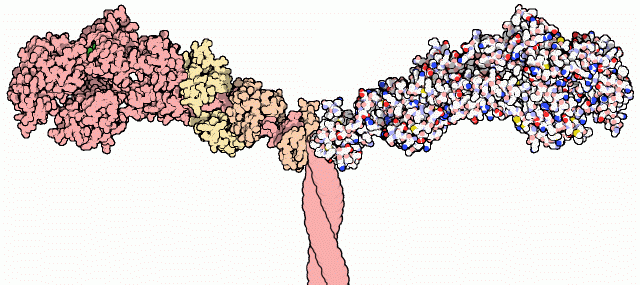 | 640×285 (46 kB) | Sysop (diskuse | příspěvky) | (Obrázek + Description: Part of the myosin structure, atoms in the heavy chain are colored red on the left-hand side, and atoms in the light chains are colored orange and yellow. (image PDB) * Source: http://www.pdb.org/ ** Houdusse, A., Kalabokis, V. N.,) |
- Editovat tento soubor v externím programu (Více informací najdete v nápovědě pro nastavení.)
Odkazy na soubor
Na soubor odkazuje tato stránka:


