V encyklopedii Allmultimedia.cz byl aktivován špičkový grafický skin Foreground.
Foreground plně podporuje – RWD, HTML 5.0, Super Galerii a YouTube 2.0 !
Foreground plně podporuje – RWD, HTML 5.0, Super Galerii a YouTube 2.0 !
Soubor:Chlamydomonas TEM 17.jpg
Z Multimediaexpo.cz
Velikost tohoto náhledu je: 751 × 600 pixelů
Obrázek ve vyšším rozlišení (rozměr: 2 450 × 1 956 pixelů, velikost souboru: 1,51 MB, MIME typ: image/jpeg)
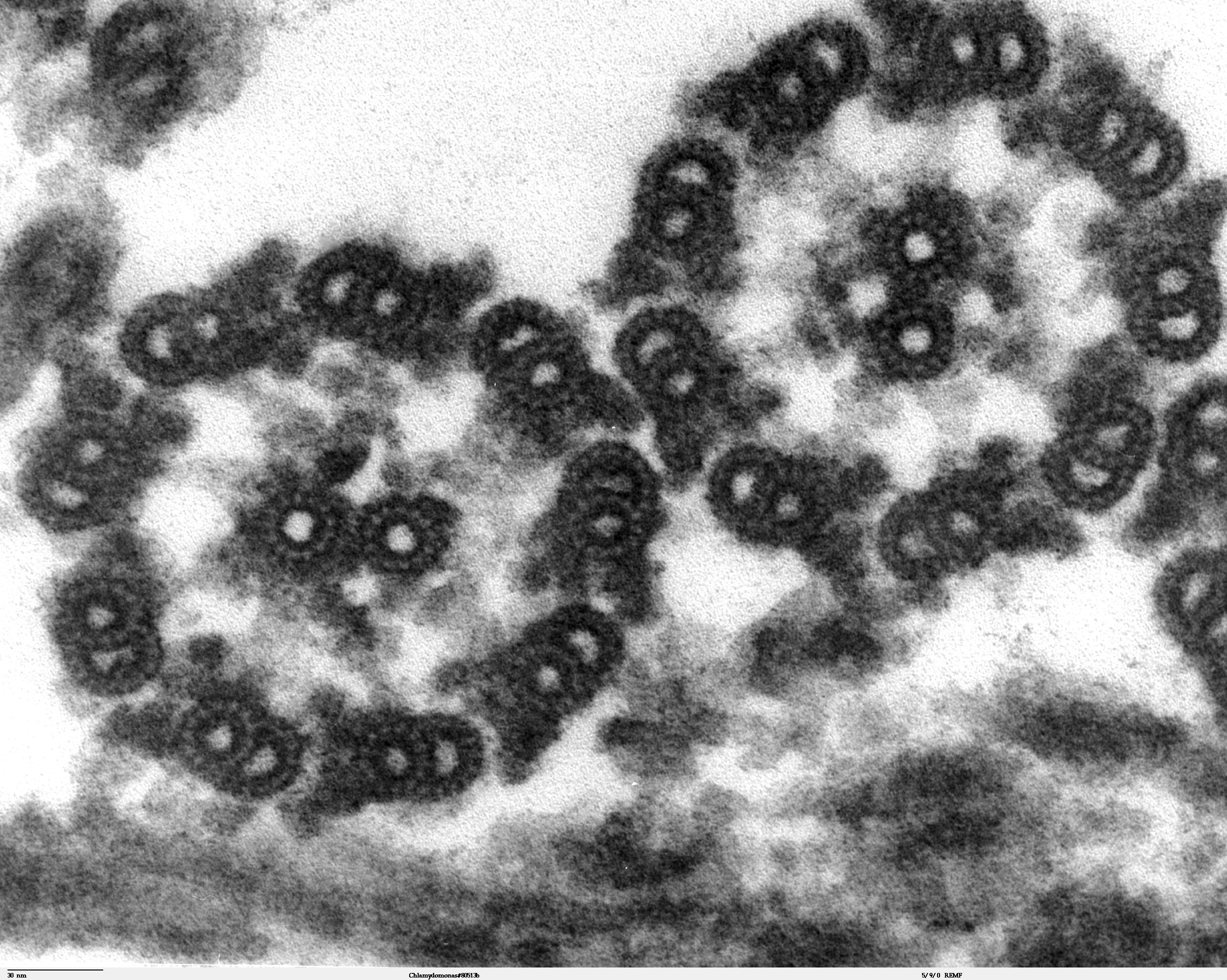
Fotografie + Description: Transmission electron microscope image, showing an example of green algae (Chlorophyta).
- Chlamydomonas reinhardtii is a unicellular flagellate used as a model system in molecular genetics work and flagellar motility studies.
- This image is a thin x-section cut through the isolated axoneme. Chlamydomonas flagella have the "9+2" structure characteristic of all eukaryotic cells. The axoneme has a central unit containing two single microtubules and nine peripheral doublet microtubules (known as the "9+2"). Dynein sidearms project from the A tubule of each doublet. Also visible in this image are the radial spokes and the inner sheath.
- Date: 20 September 2007
- Source: Source and public domain notice at http://remf.dartmouth.edu/imagesindex.html
- Author: Dartmouth Electron Microscope Facility, Dartmouth College
+ pochází z Wikimedia Commons, kde má status – This work has been released into the public domain by its author, Dartmouth Electron Microscope Facility, Dartmouth College. This applies worldwide.
Historie souboru
Kliknutím na datum a čas se zobrazí tehdejší verze souboru.
| Datum a čas | Náhled | Rozměry | Uživatel | Komentář | |
|---|---|---|---|---|---|
| současná | 15. 4. 2014, 15:36 | | 2 450×1 956 (1,51 MB) | Sysop (diskuse | příspěvky) | (MW1.15-Fotografie) |
- Editovat tento soubor v externím programu (Více informací najdete v nápovědě pro nastavení.)
Odkazy na soubor
Na soubor odkazuje tato stránka:


