The English encyclopedia Allmultimedia.org will be launched in two phases.
The final launch of the Allmultimedia.org will take place on February 24, 2026
(shortly after the 2026 Winter Olympics).
Dovolená : 23. prosinec 2025 — 29. prosinec 2025
Holidays : December 23, 2025 — December 29, 2025
The final launch of the Allmultimedia.org will take place on February 24, 2026
(shortly after the 2026 Winter Olympics).
Dovolená : 23. prosinec 2025 — 29. prosinec 2025
Holidays : December 23, 2025 — December 29, 2025
Soubor:Histology bse.jpg
Z Multimediaexpo.cz
Větší rozlišení není k dispozici.
Histology_bse.jpg (rozměr: 700 × 558 pixelů, velikost souboru: 73 kB, MIME typ: image/jpeg)
Fotografie + Description
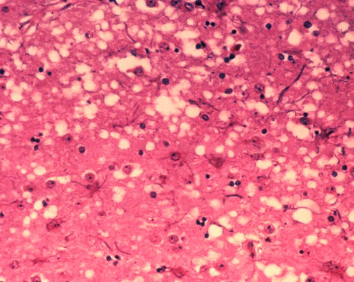
- English: This micrograph of brain tissue reveals the cytoarchitectural histopathologic changes found in bovine spongiform encephalopathy. The presence of vacuoles, i.e. microscopic “holes” in the gray matter, gives the brain of BSE-affected cows a sponge-like appearance when tissue sections are examined in the lab.
Nederlands: Deze microscopische opname toont hersenweefsel van een koe die aan BSE gestorven is. Tussen de hersencellen ziet men duidelijk verschillende vacuoles, die deze coupe (weefselsnede) een sponsachtig aanzicht geven.
- Deutsch: Das Bild zeigt die histopathologischen Veränderungen die bei einer Infektion mit BSE auftreten. Die Vakuolen, die in der grauen Substanz (substantia grisea) auftreten geben dem Bild ein schwamm-artiges Aussehen.
- Français : Cette coupe de tissu cérébral montre les modifications histopathologiques de l'organisation cellulaire lors d'une encéphalopathie spongiforme bovine. la présence de vacuoles, c'est-à-dire des "trous" microscopiques dans le tissu cérébral, donne au cerveau de vaches atteintes de l'ESB un aspect en éponge à l'examen des tissus en laboratoire.
- Date: 2003
- Source: Public Health Image Library, APHIS: http://www.aphis.usda.gov/lpa/issues/bse/bse_photogallery.html
- Author: Dr. Al Jenny
+ pochází z Wikimedia Commons, kde má status – This image or file is a work of a United States Department of Agriculture employee, taken or made as part of that person's official duties. As a work of the U.S. federal government, the image is in the public domain.
Historie souboru
Kliknutím na datum a čas se zobrazí tehdejší verze souboru.
| Datum a čas | Náhled | Rozměry | Uživatel | Komentář | |
|---|---|---|---|---|---|
| současná | 4. 11. 2013, 14:32 | | 700×558 (73 kB) | Frill80 (diskuse | příspěvky) | (FFrill200) |
- Editovat tento soubor v externím programu (Více informací najdete v nápovědě pro nastavení.)
Odkazy na soubor
Na soubor odkazují tyto 2 stránky:


