V encyklopedii Allmultimedia.cz byl aktivován špičkový grafický skin Foreground.
Foreground plně podporuje – RWD, HTML 5.0, Super Galerii a YouTube 2.0 !
Foreground plně podporuje – RWD, HTML 5.0, Super Galerii a YouTube 2.0 !
Soubor:Retina-diagram.png
Z Multimediaexpo.cz
Velikost tohoto náhledu je: 800 × 356 pixelů
Obrázek ve vyšším rozlišení (rozměr: 1 280 × 570 pixelů, velikost souboru: 212 kB, MIME typ: image/png)
Obrázek + Description:
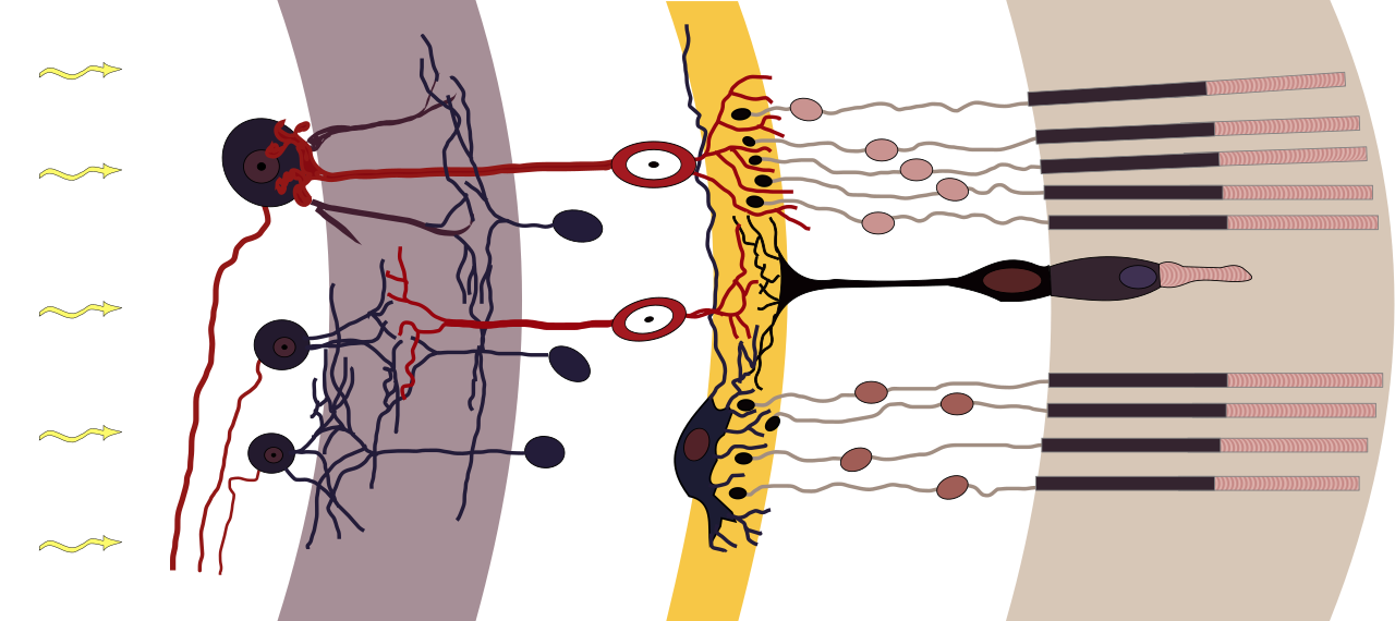
- Deutsch: Axialer Aufbau der Retina (aus Cajal, 1911). (Cajal, 1991): S. R. Y. CAJAL, Histologie Du Système Nerveux de lHomme et Des Vertébrés, Maloine, Paris, 1911
- Nervenzelltypen der Netzhaut schematisch, Licht fällt von links ein, weiß unterlegt die zellkernreichen Schichten
- v. l. n. r.: weiß: Ganglienzellen und ihre Axone, grau: Innere plexiforme Schicht, weiß: Amakrine Zellen, Bipolare, Horizontalzellen, gelb: Äußere plexiforme Schicht, weiß: Fotorezeptoren, hellbraun: Fotorezeptoren Außensegmente
- English: Axial organization of the retina (from Cajal, 1911). (Cajal, 1991): S. R. Y. CAJAL, Histologie Du Système Nerveux de lHomme et Des Vertébrés, Maloine, Paris, 1911
- Date: chris 論 12:06, 13 August 2009 (UTC)
- Source:
- Fig_retine.png
- Fig retine bended.png
- Author: Ramón y Cajal (Fig_retine.png)
- derivative work Fig retine bended.png: Anka Friedric
+ pochází z Wikimedia Commons, kde má status – This file is licensed under the Creative Commons Attribution-Share Alike 3.0 Unported license. (CC BY-SA 3.0)
Historie souboru
Kliknutím na datum a čas se zobrazí tehdejší verze souboru.
| Datum a čas | Náhled | Rozměry | Uživatel | Komentář | |
|---|---|---|---|---|---|
| současná | 21. 3. 2022, 09:36 | | 1 280×570 (212 kB) | Sysop (diskuse | příspěvky) | (Fotografie + ) |
- Editovat tento soubor v externím programu (Více informací najdete v nápovědě pro nastavení.)
Odkazy na soubor
Na soubor odkazuje tato stránka:


