The English encyclopedia Allmultimedia.org will be launched in two phases.
The final launch of the Allmultimedia.org will take place on February 24, 2026
(shortly after the 2026 Winter Olympics).
Dovolená : 23. prosinec 2025 — 29. prosinec 2025
Holidays : December 23, 2025 — December 29, 2025
The final launch of the Allmultimedia.org will take place on February 24, 2026
(shortly after the 2026 Winter Olympics).
Dovolená : 23. prosinec 2025 — 29. prosinec 2025
Holidays : December 23, 2025 — December 29, 2025
Soubor:Tuberculosis-x-ray-1.jpg
Z Multimediaexpo.cz
Větší rozlišení není k dispozici.
Tuberculosis-x-ray-1.jpg (rozměr: 590 × 542 pixelů, velikost souboru: 46 kB, MIME typ: image/jpeg)
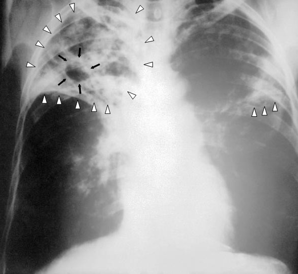
Fotografie + English: An anteroposterior X-ray of a patient diagnosed with advanced bilateral pulmonary tuberculosis. This AP X-ray of the chest reveals the presence of bilateral pulmonary infiltrate (white triangles), and „caving formation“ (black arrows) present in the right apical region.The diagnosis is far-advanced tuberculosis.
- Deutsch: Eine Röntgenaufnahme im anterior-posterioren Strahlengang eines Patienten, bei dem eine beidseitige Lungentuberkulose festgestellt wurde. Diese Thorax-Aufnahme zeigt beidseitige Lungeninfiltrate (weiße Dreiecke) und eine sogenannte „Kaverne“ (schwarze Pfeile) im rechten Oberfeld. Sie entspricht der Diagnose einer fortgeschrittenen Lungentuberkulose.
- Polski: Zdjęcie rentgenowskie w projekcji przednio-tylnej AP pacjenta ze zdiagnozowaną zaawansowaną, obustronną gruźlicą płuc. Na tym zdjęciu AP klatki piersiowej uwidoczniono obecność obustronnego zajęcia płatów (białe trójkąty) i "formowania jam" (czarne trójkąty) obecne w prawym szczycie płuca. Zdiagnozowano zaawansowną gruźlicę.
- Español: Una placa anteriorposterior de rayos X de un paciente diagnosticado con avanzado tuberculosis en ambos pulmones. Esta radiografia del pecho revela la presencia de una infiltracion (triangulos blancos), y una cavidad (flechas negras) presente en el lado derecho. El diagnostico es Tuberculosis en un grado severo de avance.
- Français : Radiographie thoracique d'un patient atteint d'une tuberculose a un stade avancé touchant les poumons de façon bilatérale. On décèle la présence de multiple micro-nodules bilatéraux a prédominance supérieure (triangles blancs) ainsi que la présence d'un caverne tuberculeux au niveau apical droit (flèches noires).
- Русский: Рентгенограмма в передне-задней проекции пациента с двухсторонним туберкулёзом лёгких. На снимке выявляется двухстороннее ограниченное затемнение (инфильтрация легочной ткани, отмечена белыми треугольными стрелками) и полостное образование (формирующаяся каверна, отмечена чёрными стрелками) в верхем поле правого легкого. Диагноз: прогрессирующий туберкулёз.
- Italiano: Radiogramma del torace in proiezione anteroposteriore in paziente con tubercolosi polmonare bilaterale avanzata. Presenza di infiltrati bilaterali (triangoli bianchi} e formazioni cavitarie (frecce nere) in apice polmonare destro. La diagnosi è tubercolosi in fase avanzata
- Date: 1972
- Source: This media comes from the Centers for Disease Control and Prevention's Public Health Image Library (PHIL), with identification number #2543.
- Author: Unknown
+ pochází z Wikimedia Commons, kde má status – This image is a work of the Centers for Disease Control and Prevention, part of the United States Department of Health and Human Services, taken or made as part of an employee's official duties. As a work of the U.S. federal government, the image is in the public domain.
Historie souboru
Kliknutím na datum a čas se zobrazí tehdejší verze souboru.
| Datum a čas | Náhled | Rozměry | Uživatel | Komentář | |
|---|---|---|---|---|---|
| současná | 9. 4. 2014, 13:12 | | 590×542 (46 kB) | Sysop (diskuse | příspěvky) | (MW1.15-Fotografie) |
- Editovat tento soubor v externím programu (Více informací najdete v nápovědě pro nastavení.)
Odkazy na soubor
Na soubor odkazuje tato stránka:


